
外科切除可能な原発乳癌の患者予後を左右する因子の中で,最も強力なものの一つが腋窩リンパ節転移の程度(n因子またはpN因子)である。n因子が同一の乳癌であっても予後はさまざまで,n因子とは独立した癌細胞の特性を表すような予後因子の研究が行われてきた。その中で,HE標本を用いて比較的簡便に実施可能で汎用性の高い病理学的グレード分類が注目され,なかでも乳癌症例の大多数を占める浸潤性乳管癌における組織学的グレード分類(histological grading)または核グレード分類(nuclear grading)の有用性が強調されてきた1)~5)。また病理学的グレード分類は予後予測だけでなく,治療方針の決定やプロトコール研究の際の適格症例選択の目的でも用いられてきた6)7)。
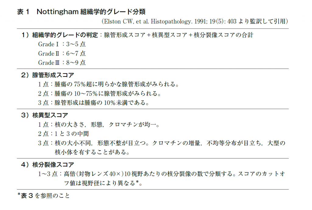
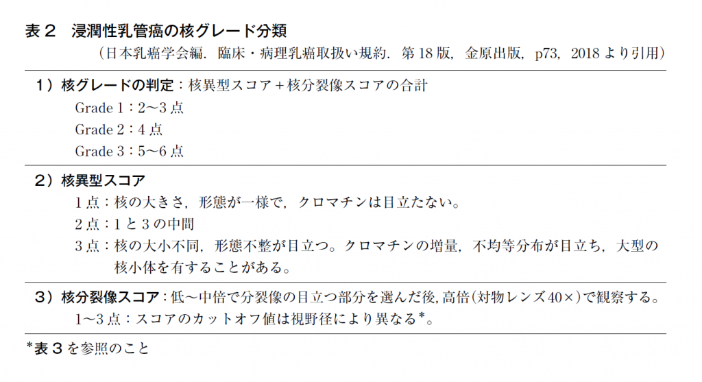
病理学的グレード分類は癌細胞の顔つきの悪さの程度を反映し,n因子や腫瘍浸潤径とは独立した予後因子である。組織学的グレード分類のうち,最も広く知られているのは英国のBloom & Richardsonによる組織学的グレード分類で,Elston & Ellisらがさらに改良を加えている(Nottingham分類:表1)3)。これらの分類では,浸潤性乳管癌の浸潤部を対象にHE染色標本を用いて判定し,腺管形成,核異型,核分裂像(各1~3点)の3項目のスコアで判定する(合計3~9点)。元来は通常型の浸潤性乳管癌に対するグレードであったが,近年では組織型にかかわらず浸潤癌に適用している報告が多い。核グレード分類もよく用いられており,Blackらの分類4)やLe Doussalらの分類5),および「乳癌取扱い規約」の核グレード分類7)がある(表2)。「乳癌取扱い規約」の核グレード分類では核異型(nuclear atypia),核分裂像の数(mitotic count)の2項目をそれぞれ1~3点にスコア化し,両者を足して判定している(合計2~6点)。組織学的グレード分類,核グレード分類のいずれも最終的に三段階に分けられており,低異型度のグレード1の乳癌は予後良好で,グレード2は中間,高異型度のグレード3の乳癌は予後不良である1)~4)8)~10)。
一方で,病理学的グレード分類は,主観的で再現性が低いことが指摘されてきた。客観性・再現性向上のため,複数の病理医が参加し,同一の組織標本を対象としてグレード分類を行う検討がなされている4)6)7)。わが国ではNSAS―BC試験に用いる核グレードの診断者間一致率の向上を目的とし,50人以上の病理医が参加したスライドカンファレンスが5回にわたって開催された実例がある11)。同検討は,診断者間一致率が80%を超える症例が,回を重ねるごとに増加すること(33→55%),診断者間一致率が低い症例は,同一病理医の診断再現性も悪いことを明らかにしている。

参考文献
1)Bloom HJ, Richardson WW. Histological grading and prognosis in breast cancer;a study of 1409 cases of which 359 have been followed for 15 years. Br J Cancer. 1957;11(3):359―77. [PMID:13499785]
2)Freedman LS, Edwards DN, McConnell EM, Downham DY. Histological grade and other prognostic factors in relation to survival of patients with breast cancer. Br J Cancer. 1979;40(1):44―55. [PMID:454563]
3)Elston CW, Ellis IO. Pathological prognostic factors in breast cancer. I. The value of histological grade in breast cancer:experience from a large study with long―term follow―up. Histopathology. 1991;19(5):403―10. [PMID:1757079]
4)Cutler SJ, Black MM, Friedell GH, Vidone RA, Goldenberg IS. Prognostic factors in cancer of the female breast. II. Reproducibility of histopathologic classification. Cancer. 1966;19(1):75―82. [PMID:5901409]
5)Le Doussal V, Tubiana―Hulin M, Friedman S, Hacene K, Spyratos F, Brunet M. Prognostic value of histologic grade nuclear components of Scarff―Bloom―Richardson(SBR). An improved score modification based on a multivariate analysis of 1262 invasive ductal breast carcinomas. Cancer. 1989;64(9):1914―21. [PMID:2551477]
6)Dalton LW, Page DL, Dupont WD. Histologic grading of breast carcinoma. A reproducibility study. Cancer. 1994;73(11):2765―70. [PMID:8194018]
7)Tsuda H, Akiyama F, Kurosumi M, Sakamoto G, Watanabe T. Establishment of histological criteria for high―risk node―negative breast carcinoma for a multi―institutional randomized clinical trial of adjuvant therapy. Japan National Surgical Adjuvant Study of Breast Cancer(NSAS―BC)Pathology Section. Jpn J Clin Oncol. 1998;28(8):486―91. [PMID:9769782]
8)Bult P, Manders P, Straatman HM, Tjan―Heijnen VC, Beex LV, Hendriks J, et al. In primary breast cancer the mitotic activity yields similar prognostic information as the histological grade:a study with long―term follow―up. Breast Cancer Res Treat. 2010;122(1):77―86. [PMID:19760038]
9)Ono M, Tsuda H, Yunokawa M, Yonemori K, Shimizu C, Tamura K, et al. Prognostic impact of Ki―67 labeling indices with 3 different cutoff values, histological grade, and nuclear grade in hormone―receptor―positive, HER2―negative, node―negative invasive breast cancers. Breast Cancer. 2015;22(2):141―52. [PMID:23584595]
10)Schwartz AM, Henson DE, Chen D, Rajamarthandan S. Histologic grade remains a prognostic factor for breast cancer regardless of the number of positive lymph nodes and tumor size:a study of 161 708 cases of breast cancer from the SEER Program. Arch Pathol Lab Med. 2014;138(8):1048―52. [PMID:25076293]
11)Tsuda H, Akiyama F, Kurosumi M, Sakamoto G, Watanabe T. The efficacy and limitations of repeated slide conferences for improving interobserver agreement when judging nuclear atypia of breast cancer. The Japan National Surgical Adjuvant Study of Breast Cancer(NSAS―BC)Pathology Section. Jpn J Clin Oncol. 1999;29(2):68―73. [PMID:10089946]